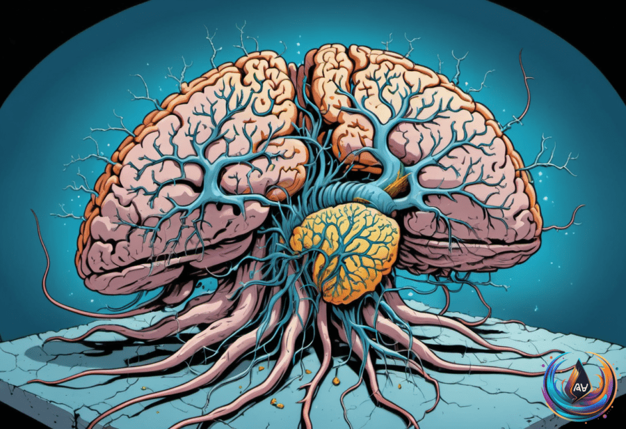
A hibernation-like state can erase more than half of a mouse’s synapses, yet the animals still retain their memories. When the mice wake, most of those lost connections return, and 82 percent reappear in the same spot.
The finding creates a puzzle for neuroscience. Synapses are connections between neurons, and one view of memory holds that memory traces depend on the strength of individual synapses. Kazumasa Tanaka, a neuroscientist at the Okinawa Institute of Science and Technology Graduate University, described the expected result plainly: “If you accept that memory traces reside in the efficacy of individual synapses, if you lose more than half of the synaptic connections, of course what you’d expect is impairment of the memory afterwards.”
A dramatic reset that does not erase memory
The mice spent 48 hours in a hibernation-like state. During that time, synaptic activity dropped by about 70 percent once hibernation set in, and the animals experienced a major loss of synapses. Despite that loss, they retained their memories after hibernation.
Takeshi Sakurai, a neuroscientist at the University of Tsukuba, explained how the state was induced: “With our protocol, we can bring mice’s body temperature down to somewhere around 20° Celsius, and their heart rate and breathing rate decrease significantly as well.” The change affects the body and the brain at the same time, creating a state that resembles hibernation.
After arousal, the vanished synapses reappeared. The fact that 82 percent returned to the same spot suggests that the connections did not simply grow back in random locations. Their return followed a pattern, even though the mice had passed through a state that removed more than half of their synapses.
The result leaves memory and synapse strength connected, but not in a simple way. The mice remembered tasks even after the large drop in synaptic connections, showing that memory can survive a major change in the brain’s physical links.
Sleep depends on more than one brain pathway
Other work in mice has focused on the neurons that control sleep itself. Activating neurons in the median raphe nucleus increases sleep duration and quality, while inhibiting these neurons decreases sleep by about 70 percent. When sleep was inhibited, mice spent about 6.5 extra hours awake each day.
Will Joo, a researcher at the University of Basel, said of these neurons: “They are crucial to promote sleep, and may be key components of the neural circuitry that generates sleep.” The results point to an important role for the median raphe nucleus, but they do not make it the brain’s only sleep switch.
Luis de Lecea, a researcher at Stanford University, placed that pathway in a wider picture: “The effect of this major neuronal path on sleep is very strong, but, as we have learned since we started manipulating neuronal circuits, each path provides nuances. The brain has many ways to wake up and fall asleep.”
That complexity also shows up during sleep deprivation. In a separate line of research, small clusters of neurons can enter a local sleep state for a few hundred milliseconds while the rest of the brain remains awake. Local sleep can reduce markers of synaptic strength in specific brain regions, linking brief rest in small neural groups with changes in their connections.
When the awake brain takes short rests
Local sleep is more likely when the brain is overused or deprived of sleep. Highly demanding cognitive tasks can raise the chance of local sleep in the brain regions handling that work, suggesting that neural circuits may need short breaks after sustained effort.
Thomas Andrillon, a neuroscientist at the Paris Brain Institute, described this pressure as both time-based and use-based: “It’s not just time-dependent – so how much you have slept – it’s also use-dependent.” In other words, the need for rest depends not only on how long an animal has stayed awake, but also on how much particular brain areas have been used.
Low-arousal states during mindless tasks may create the right conditions for these brief local pauses. Andrillon explained, “It makes sense that, in this situation, where monitoring the outside is not that interesting, your brain could use these opportunities to do a bit of housekeeping.”
That housekeeping may help explain why wakeful rest can support thinking. Local sleep induced after learning helped mice remember tasks as well as mice that took a nap, and wakeful rest may restore cognitive functions in a way that resembles sleep.
Researchers including Giulio Tononi and Chiara Cirelli of the University of Wisconsin-Madison, Alex Schier and Will Joo of the University of Basel, Angelica Quercia of the Oasi Research Institute-IRCCS, and Mark Wu of Johns Hopkins University have contributed to work examining sleep, neural activity, and memory. Together, these findings present the sleeping and waking brain as flexible rather than passive.
The studies do not describe one single process. Hibernation-like states remove many synapses and allow most to return; targeted neuron activity can change how long mice sleep; and brief local sleep can appear inside an otherwise awake, sleep-deprived brain. The common thread is that the brain can alter its connections and activity without simply losing the memories or functions those systems support.
Based on