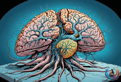
The brain may be doing far more than sending signals from one point to another. New findings reveal waves that travel across the cortex, reorganize activity in real time, and expose how information moves through neural tissue.
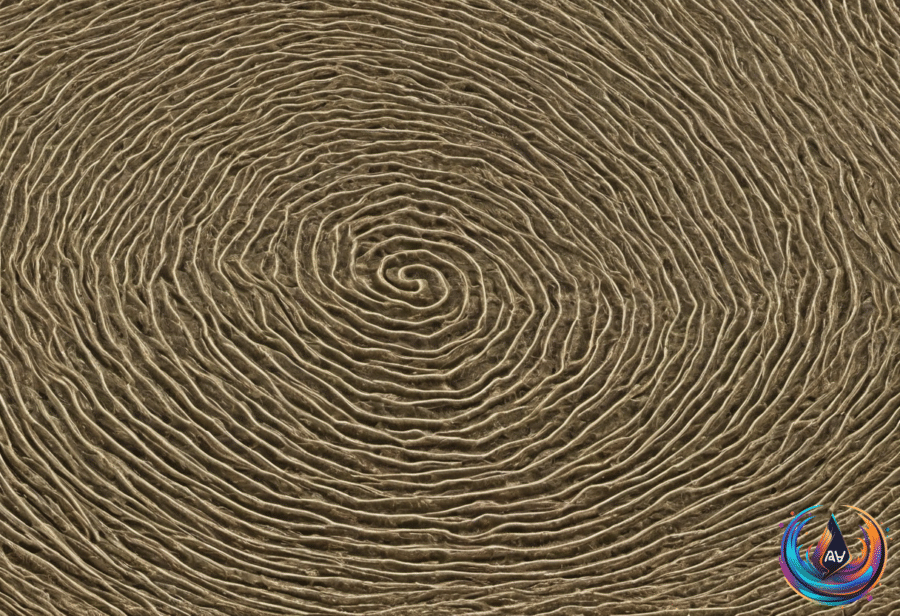
These waves are not simple ripples. They include source waves, sink waves, and vortexlike spiral waves, with different patterns linked to different behavioral tasks. Together, the results point toward a brain whose activity is constantly shifting across space, not merely switching on and off in isolated regions.
A Moving Map of Cortical Activity
Traveling waves can show the direction of information propagation in the brain, giving researchers a way to track activity as it moves through the cortex. That direction may reveal what the brain is doing during a task, even when the underlying neural signals are difficult to interpret.
Earl K. Miller, a cognitive neuroscientist at the Massachusetts Institute of Technology, described the field’s changing focus: “The work coming out is moving this from ‘Are they relevant?’ to ‘This is a major motif of how the cortex processes information,’”
The findings also suggest that wave patterns change with behavior. A task can produce one traveling pattern, while another task produces a different pattern, creating a dynamic link between neural motion and what a person is doing.
Joshua Jacobs from the University of Chicago offered another way to understand the result: “Even if the traveling wave was just the result of neurons firing, like the sound of the engine, it still tells us something very interesting about the brain based on what the person is doing.”
That idea gives the waves a powerful role even when they are not treated as the direct cause of brain activity. Their shape and direction can still carry information about behavior and neural organization.
Shared Dynamics Beneath Anesthesia
Another set of findings examines neural activity during anesthesia and finds conserved dynamic signatures across species. The shared patterns suggest that some features of brain dynamics remain recognizable even when conscious experience is altered.
George A. Mashour, an author and researcher, and Zirui Huang, an author and researcher, are among the named figures connected to this area of work. Uma Mohan, a neuroengineer at the National Institutes of Health, also appears among the researchers associated with these advances.
The cross-species result adds a new layer to the study of brain dynamics. Traveling waves describe activity moving across the cortex, while conserved signatures during anesthesia point to patterns that can persist across different species. Both findings shift attention toward the motion and structure of neural activity.
MEG Reaches Into Cortical Layers
High-density MEG is also opening a path toward laminar inference, the process of estimating the cortical depth most consistent with measured signals. A multilayer framework can estimate where activity sits within the cortex, reaching beyond a broad surface-level picture.
The limits are precise. Laminar inference is feasible at −50 to −35 dB SNR, while fine-scale inference becomes possible at −20 dB and above. At −50 dB SNR, inference remains at chance for middle laminae but performs significantly better for superficial and deep laminae.
- Accurate laminar inference requires a co-registration error of 2 mm or less.
- Reconstruction accuracy improves with higher SNR and smaller co-registration errors.
- Small misalignments of ≤2 mm have minimal effects on accuracy.
- Errors exceeding ~3 mm become unreliable.
- At errors of 5 mm, only deep and superficial sources retain some discriminability.
Error was lowest at 0 mm co-registration error, with a permutation test showing a standardized difference of −4.84 and p 0.06. At very low SNR levels of −100 dB and below, reconstruction error was indistinguishable from chance.
The framework also accounts for regional variability in cortical laminar thickness. At sufficiently high SNR, true laminar inference remains possible even when that variation is included, showing that the method can preserve depth information under more realistic conditions.
Speech Imagery Nearly Matches Speaking
Neural dynamics also connect imagined speech with physical articulation. A neural architecture for speech imagery and articulation uses somato-cognitive organization, linking supramodal planning with modality-specific representations.
The prediction results close the gap between imagining speech and producing it. Across nine participants, labeled S1–S9, speech imagery reached a median prediction accuracy of 80.4%, compared with 78.3% for speech articulation.
That close match matters because it shows that imagined speech can produce neural patterns with prediction accuracy comparable to actual articulation. The brain’s planning systems and speech-specific representations remain linked across both conditions.
Across these findings, one message keeps gaining force: neural activity is organized through movement, depth, shared dynamics, and behavior-linked patterns. Traveling waves can reveal direction, MEG can estimate cortical layers, anesthesia can preserve conserved signatures across species, and speech imagery can approach the neural predictability of speaking.
The next stage will bring these views together. The brain is not only a network of connected regions; it is a shifting landscape where waves travel, layers separate signals, and thought can leave a measurable path.
Based on
- Surprisingly Complex Waves Reveal the Brain’s Inner Workings — quantamagazine.org
- Conserved neural dynamics of anesthesia and the oblivion of species | Nature Neuroscience — nature.com
- Multilayer MEG source modelling enables depth-resolved laminar inference in humans – Nature Communications — nature.com
- A neural architecture for imagined and overt speech motor dynamics | Nature Neuroscience — nature.com